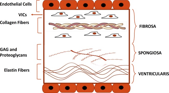
Figure 1.
Histological structure of the aortic valve. Depicted is the histological structure of the healthy aortic valve. Fibrosa, spongiosa, and, ventricularis are the three layers that make up the structure of a normal aortic valve. The Fibrosa layer is composed of type I and III collagen fibers and contains also VICs. Spongiosa and ventricularis layers are respectively composed of GAG and proteoglycans and elastin fibers. Endothelial cells form a monolayer on each side of the cusp. GAG, glycosaminoglycans; VIC, valve interstitial cell.

