Figure 2.

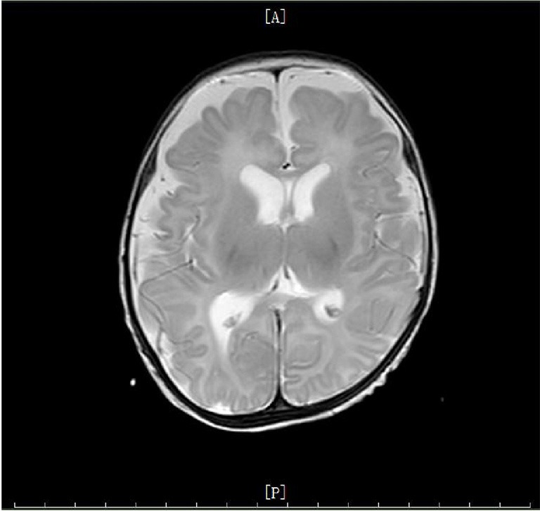
Cranial MRI imaging of the patient. The lateral frontotemporal parietal space on both sides was widened, and sulcus fissure was widened and increased. All of these suggested a possibility of brain dysplasia.

Cranial MRI imaging of the patient. The lateral frontotemporal parietal space on both sides was widened, and sulcus fissure was widened and increased. All of these suggested a possibility of brain dysplasia.