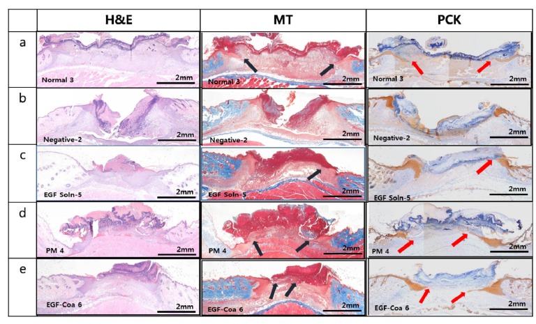
Figure 4.
Histological images of wound beds with hematoxylin and eosin (H&E), Masson’s trichrome (MT), and pan-cytokeratin (PCK) staining. (a) Normal (non-diabetic mice), (b) Negative (diabetic mice) treated with PBS, (c), (d), and (e) Diabetic mice treated with EGF solution, EGF-PM, and EGF-Coa. Black (MT) and red (PCK) arrows indicate the pointed fronts of horizontal migration of keratinocytes along the surface of newly formed granulation tissue.

