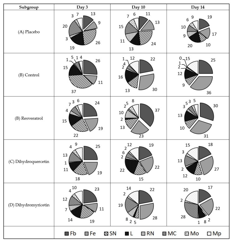
Figure 6.
Diagram of cell types’ share in the cell repertoire of the wound defect surface is shown in the wounds infected with P. aeruginosa, %. Cell types are denoted as follows: Fb—fibroblast; Fc—fibrocyte; SN—polymorphonuclear neutrophil; L—lymphocyte; RN—immature neutrophil; MC—mast cell; Mo—monocyte; Mp—macrophage. Total share of resident cells (Fb + Fc) is shown in box beneath respective sectors.

