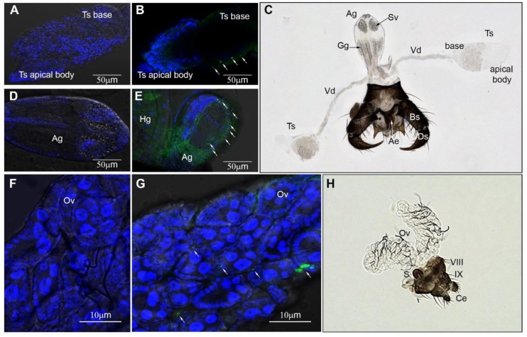
Figure 5.
Immunofluorescent VSV-staining of intrathoracically infected C. sonorensis. (A) Testis (Ts) and (D) accessory gland (Ag) dissected from non-infected (negative control) males. (B) Testis (Ts) and (E) accessory gland (Ag) dissected from males 4 days post inoculation. Arrows denote VSV-positive staining (FITC-green puncta) in the epithelial layer of the testis base and outer epithelial layer of the Ag. Cellular nuclei were stained with DAPI (blue). (C) Male reproductive anatomy (brightfield 200×). Abbreviations: Ae: aedeagus, Ag: accessory gland, Bs: basistyle, Ds: dististyle, Gg: glutinous gland, Sv: seminal vesicle, Ts: testis, Vd: vas deferens. (F) Ovaries (Ov) dissected from non-infected (negative control) females. (G) Ovaries (Ov) dissected from females 4 days post inoculation. Arrows denote VSV-positive staining (FITC-green puncta) with DAPI nuclear stain (blue). (H) Female reproductive anatomy (brightfield 200×). Abbreviations: VIII: 8th abdominal segment, IX: 9th abdominal segment, Ce: cerci, Ov: ovary, S: spermatheca.

