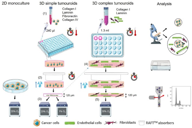
Figure 1.
Schematic of tumouroid manufacture. (1) Cancer cell lines are embedded in collagen I hydrogels in 96-well plates containing collagen IV, laminin and fibronectin. (2) Interstitial fluid is removed using commercial absorbers (RAFTTM) to create a dense cancer mass. (3) The resulting cancer mass–only tumouroid is cultured and treated with drugs. (4) For complex tumouroid manufacture, the cancer-only simple tumouroid is nested in another hydrogel containing human dermal fibroblasts (HDFs) and human umbilical vein endothelial cells (HUVECs) in 24-well plates and compressed using commercial absorbers (RAFTTM) to create a dense complex tumouroid. (5) Complex tumouroids are cultured and treated with drugs. Analysis is done using fluorescence imaging, cell viability, ELISA and cell cycle analysis using flow cytometry. Schematic is based on previous works23,26,27 and designed using SMART – Servier Medical ART.

