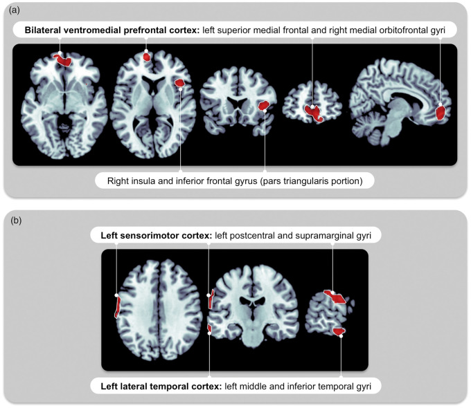
Figure 1.
Daytime brain perfusion patterns associated with OSA during REM versus NREM sleep. In the complete sample representing all OSA severities (n = 96, AHI from 0 to 97, model 1), reduced daytime rCBF is associated with (a) a higher REM-AH in the bilateral ventromedial prefrontal cortex and right insula extending to the frontal cortex; (b) a higher NREM-AH in the left sensorimotor and lateral temporal cortex. Regressions model were between REM-AH and daytime rCBF, adjusted for age, total sleep duration and NREM-AH (a); and between NREM-AH and daytime rCBF, adjusted for age, total sleep duration and REM-AH (b). Statistical analyses were performed on every voxel of gray matter. Significant regions of abnormal rCBF were identified when single voxels showed a significant regression with the OSA variable (statistical threshold at p < 0.001) and when these were surrounded by a cluster of ≥100 voxels with rCBF values that behave similarly. Similar regions of daytime hypoperfusion were observed when REM-AHI and NREM-AHI were used instead of REM-AH and NREM-AH. rCBF: regional cerebral blood flow; NREM: non-rapid eye movement sleep; REM: rapid eye movement sleep; AH: apneas + hypopneas; AHI: apnea–hypopnea index; OSA: obstructive sleep apnea.

