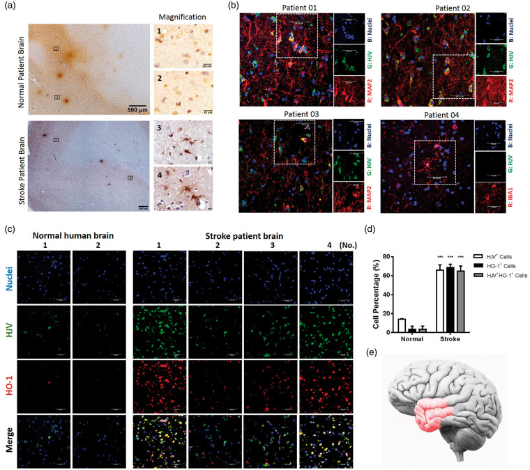
Figure 1.
Cells immunoreactive to HJV were found in human brain tissues and were more abundant in stroke patients (n = 10) than controls (n = 2). (a) Immunohistochemical staining of HJV from cortex region of normal (n = 2) and acute stroke patient (n = 10). (b) Characterization of cells expressing HJV in the stroke patient brain section (n = 4) by double immunofluorescence with anti-MAP2 or anti-IBA1 antibodies. (c) Double immunofluorescence micrographs of HJV and HO-1 from cortex region of normal (n=2) and acute stroke patients (n = 4). Scale bars: 50 μm. (d) Percentage of HJV-positive cells, HO-1-positive cells or double-positive cells among total cells from biopsy specimen. Data were expressed as mean±SD; ***, P<0.001 vs. control group. (e) Schematic of the clinical human brain tissue in reference to the infarction region (Red color).

