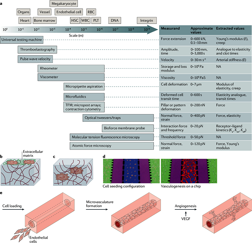
Figure 1: Tools to measure and recreate the mechanical properties of blood tissues.
a) The schematics compares the length scale of blood cells and tissues with the resolution of techniques for the investigation of the mechanical properties of blood cells and tissues. Typical measurements, values, and extracted values are also shown. b) Schematic of a 2D cell culture on a protein-conjugated hydrogel surface. c) Schematic of a 3D cell culture. Cells are encapsulated inside a hydrogel and can interact with the surrounding environment. d) Hydrogels can be used for the engineering of microvasculature by vasculogenesis (green: fibrin gels embedded with perivascular cells; purple: fluidic channels; blue: fibrin gels embedded with endothelial cells; red: microvascular network formed via vasculogenesis208. e) Hydrogels can also be applied for the engineering of microvasculature by angiogenesis, using vascular endothelial growth factor (VEGF)209.

