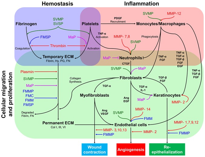
Figure 1.
Simplified diagram of the interactions between different cell types during wound healing, the contribution of MMPs and proposed wound healing mechanisms of SPs. Skin injury repair begins with hemostasis, a process which stops blood loss and provides a temporary matrix facilitating further steps in wound healing. Fibrin-rich ECM formation stimulates neutrophil-activated monocyte recruitment through TNF-α and PDGF. Both neutrophils and monocytes produce several growth factors, such as TNF-α, TGF-α, TGF-β, EGF and FGF, to enhance migration and proliferation of fibroblasts, endothelial cells, and keratinocytes to the site of injury. Fibroblasts stimulate other cells to produce collagen deposits in the ECM, wound contraction, angiogenesis and re-epithelization. Studies suggest that SPs, such as FMC, FMMP, FMM, FMSP, MaP, SVMP and SVSP, may behave similarly to endogenous MMPs during these stages. Ang, angiopoietin; CTGF, connective tissue growth factor; Col, collagen; ECM, extracellular matrix; EGF, epidermal growth factor; FGF, fibroblast growth factor; FMC, fish mucus cathepsin; FMMP, fish mucus matrix metalloprotease; FMM, fish mucus meprin; FMSP, fish mucus serine protease; FN, fibronectin; Hy, hyaluronan; IL-1, interleukin-1; MaP, maggot protease; MMP, matrix metalloproteinase; PDGF, platelet-derived growth factor; PG, proteoglycan; SVMP, snake venom metalloprotease; SVSP, snake proteinase; TGF, transforming growth factor; TNF-α, tumor necrosis factor-α; VEGF, vascular endothelial growth factor.

