
Fig. 4. The ADMIDAS cation is not present in αvβ8.
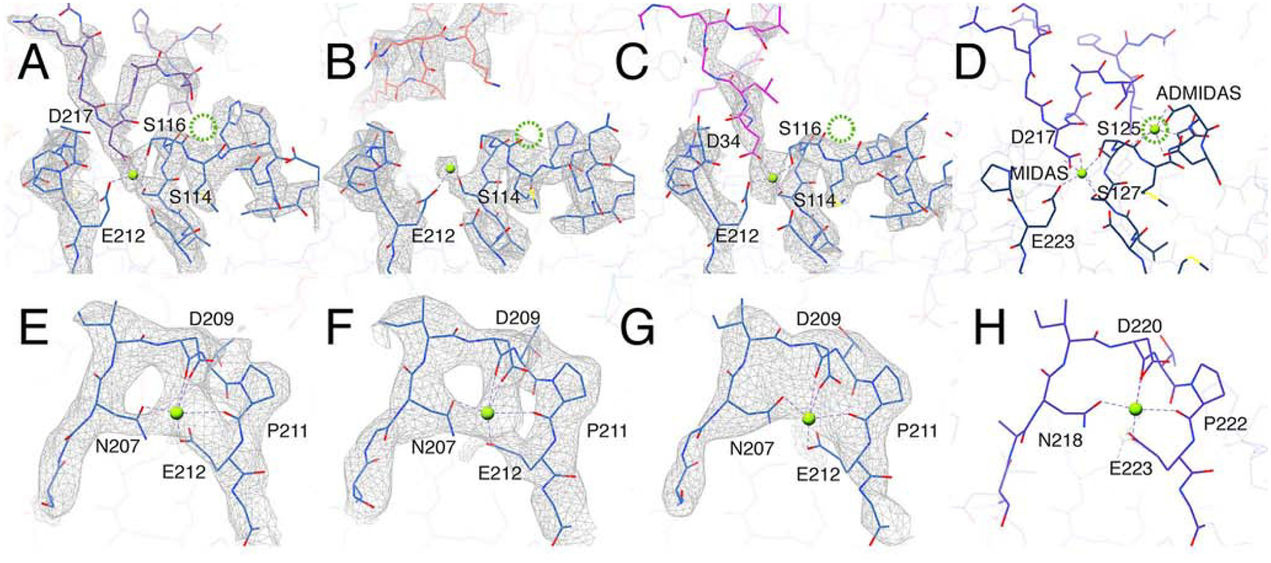
(A-C) The MIDAS cation binding site formed in αvβ8 integrin (blue) bound with: L-TGF-β1 (purple, A) C6D4 (coral, B) and C6-RGD3 (magenta, C). In all three structures there is clear density for the MIDAS cation (bright green). No density for the ADMIDAS cation is observed (the expected position for ADMIDAS is noted by the dotted green circle). Shown are the cation-coordinating residues of the integrin b subunit or the Asp of RGD of L-TGF-β (D217) or C6-RGD3 (D34). (D) The same binding interface in the αvβ6/LTGFβ crystal structure (5FFO (Dong et al., 2017)). (E-H) Maps and models of the SyMBS cation in αvβ8/L-TGF-β (E), αvβ8/C6D4 (F), αvβ8/C6-RGD3 (G), and αvβ6/LTGFβ crystal structure (5FFO) (H). All panels are color coded as in Fig. 2. Related information is in Fig. S3.