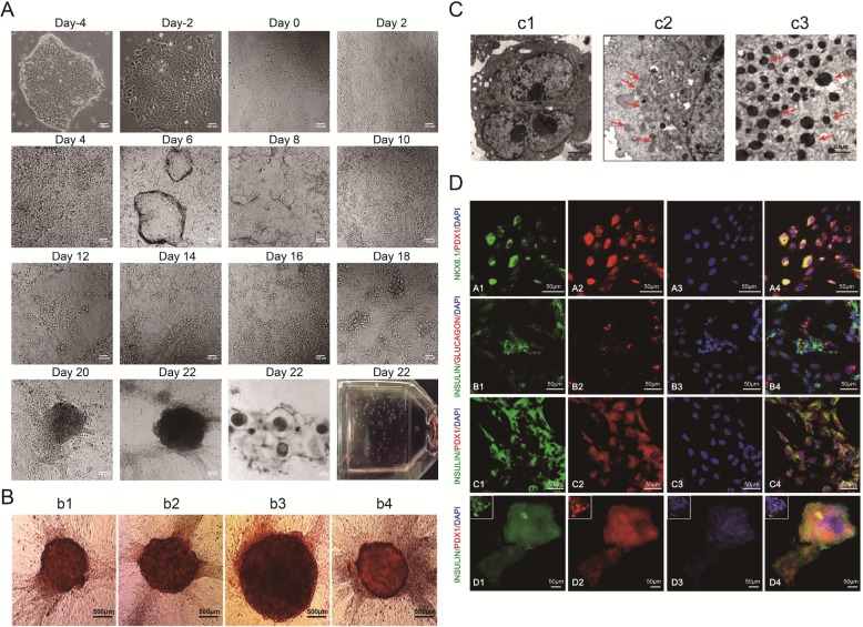
Fig. 1.
Morphology and phenotype characteristics of pancreatic β cells derived from hiPSCs at the final maturation stage. A Morphological changes of hiPSCs during differentiation into mature pancreatic β cells. B The pancreatic β cells derived from hiPSCs were stained with DTZ. Scale bars, 500 μm (b1, b2, b3, b4). C Scanning electron microscopy of IPCs derived from hiPSCs. The IPCs have secretory granules and complete capsules. Scale bars, 1 μm (c1); 0.5 μm (c2, c3). D Immunofluorescence staining showing that the differentiated hiPSCs at the final mature stage co-expressed PDX1 and NKX6.1, insulin and glucagon, and PDX1 and insulin (scale bars = 50 μm)

