Fig. 1.
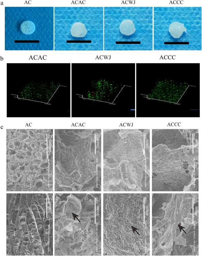
Hyaline cartilage-like gross morphology and directional distribution of cells and ECM after co-culture in ACECM-oriented scaffold. a After culture for 3 weeks, the 3D ACECM-oriented scaffolds were filled with cells and extracellular matrix in the ACAC, ACWJ and ACCC groups, and the appearance of the engineered tissue was semitransparent and glossy. The scale of black bar is 1 cm. b Dead/live staining under 3D confocal fluorescence microscopy. The 3D-oriented scaffolds are mainly filled with green fluorescent cells (living cells), with just a few red fluorescence cells (dead cells) present after cultivation for 3 days. c SEM results. The upper row shows the high-power (HP) field, and the lower row shows the low-power (LP) field; AC group, the upper and lower pictures are cross-sectional and sagittal plane views, respectively. In the ACAC, ACWJ and ACCC groups, the HP field shows cells distributed in holes, and the LP field shows the topological structure of the ECM; black arrows indicate the topological microstructure of ECM secreted by cells

