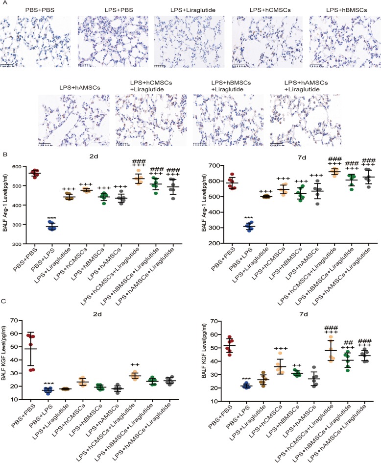
Fig. 7.
Expression of SPC, Ang-1, and KGF in ALI models. a The expression of SPC in lung sections after 7d of LPS stimulation by immunohistochemistry. Brown intracellular precipitation represents SPC protein. The expression of Ang-1 (b) and KGF (c) in BALF after LPS stimulation for 2d and 7d were detected by ELISA assay. Each group contains 6 mice. Error bars represent mean ± SD from three independent experiments. Scale bars, 50 μm. Compared with the PBS + PBS group, ***P < 0.001; compared with the LPS + PBS group, ++P < 0.01 and +++P < 0.001; compared with the corresponding MSC group, ##P < 0.01 and ###P < 0.001

