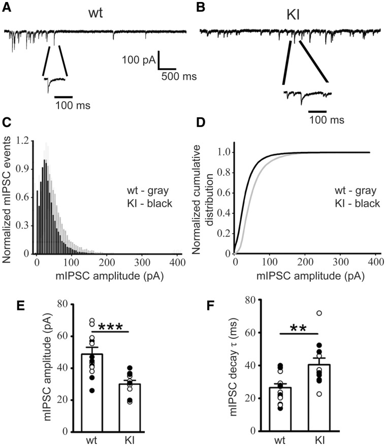
Figure 6.
mIPSCs recorded from KI mouse SS cortex layer V/VI neurons were altered. mIPSCs were recorded from SS cortex layer V/VI neurons (voltage-clamped at −60 mV with equal chloride concentration inside and outside cells) in thalamocortical slices from littermate (A) wild type and (B) KI mice. (C) Normalized mIPSC events for wild type and KI mice showed the frequency of mIPSCs in each amplitude bin. (D) Normalized cumulative distribution was plotted for wild type and KI mouse mIPSCs. Compared with mIPSCs recorded from wild type littermate mice, those from SS cortex layer V/VI neurons in KI mice had (E) significantly reduced amplitudes (wild type −49.67 ± 2.71 pA; KI −31.48 ± 1.76 pA) and (F) slowed mIPSC decay (wild type 25.05 ± 2.87 ms; KI 39.13 ± 2.72 ms). wild type, n = 6 mice; KI, n = 5 mice. Student’s t-test, **P < 0.01, ***P < 0.001. SS: somatosensory.

