
Figure 6:
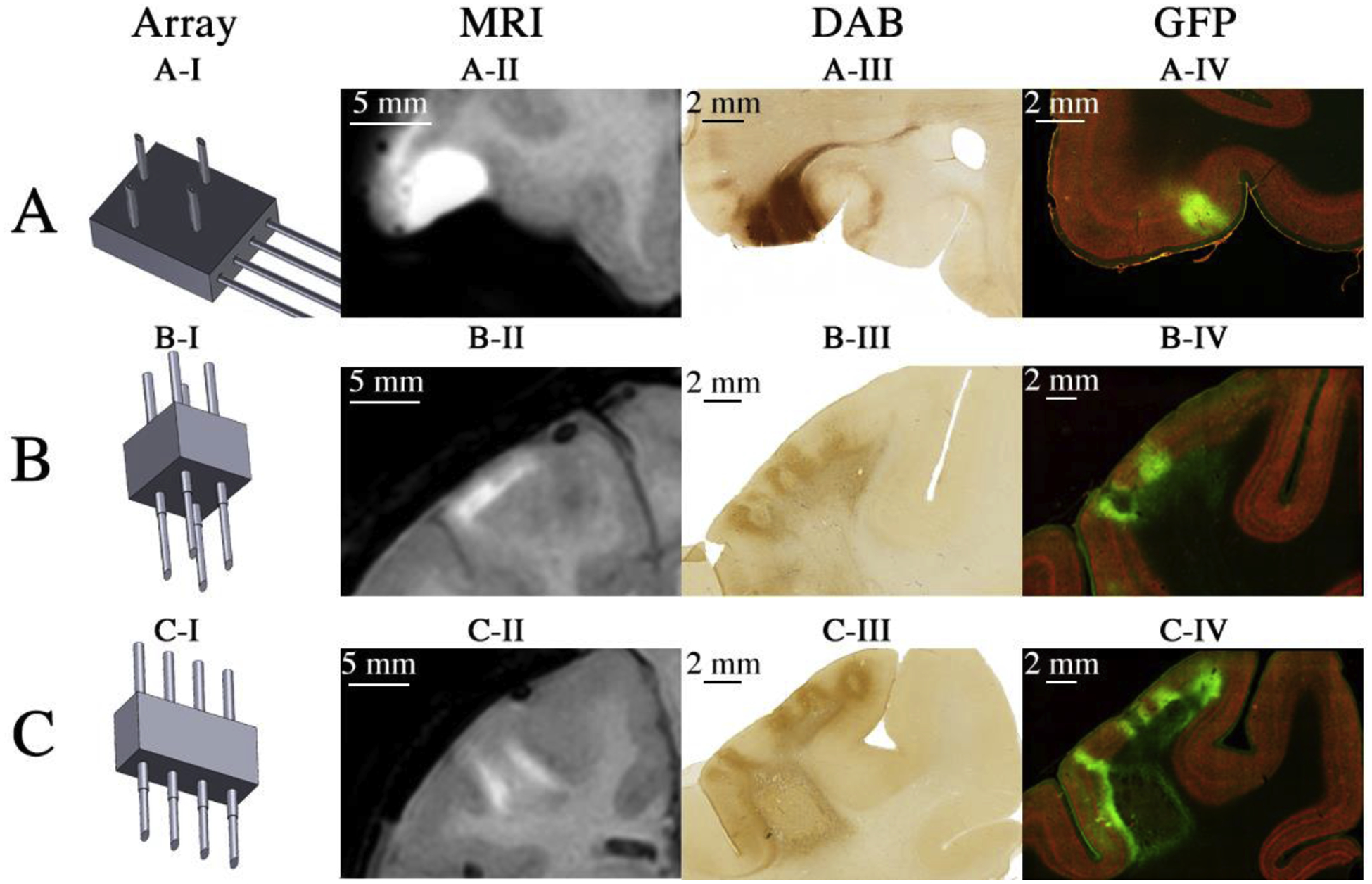
In vivo and ex vivo visualization of cortical viral injection/expression using injector arrays. (AI) Schematic representation of injector type used. (AII) Skull-stripped MR image of Monkey A acquired after virus co-infused with 5 mM Mn2+ into area 12 of ventrolateral prefrontal cortex (vlPFC). (AIII) Bright field visualization of CFP expression. (AIV) Fluorescent imaging of CFP expression. (BI – BIV) Same conventions as ‘A’, but illustrating the results of injections into primary somatosensory cortex (S1) using the needle array for surface injections of dorsal cortex of Monkey B using 0.1 mM Mn2+. (CI – CIV) Same conventions as ‘A’, but illustrating the results of two injections, one into central sulcus and one into intraparietal sulcus of Monkey B using the needle array for sulcal injections with 0.1 mM Mn2+.