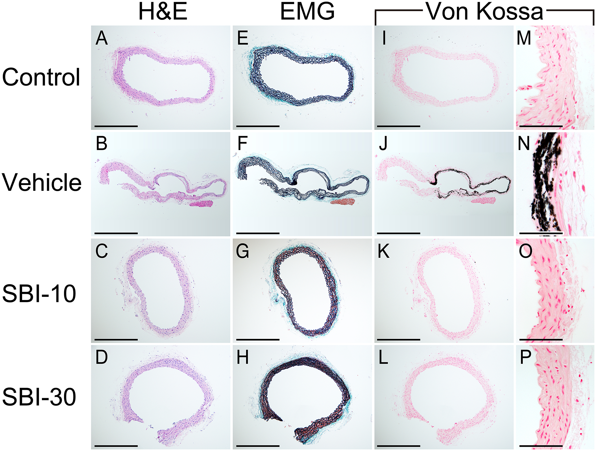
Figure 4. Micrographs of H&E, EMG, Von Kossa and Alizarin Red stained sections of the abdominal aorta.

Sections of the abdominal aorta for the Control, Vehicle, SBI-10 and SBI-30 groups are shown. Medial arterial calcification (MAC) were observed in vehicle mice as von-Kossa staining positive legions (J, N). MAC was barely observed among mice in the Control, SBI-10, and SBI-30 groups (I, K, L, M, O, P). Elastin layers in these calcified areas grossly appeared to be disorganized and disrupted (F), while no evidence of cartilaginous metaplasia, inflammation or atherosclerotic lesions were observed (B, F), in line with control samples (A, E). Scale bar, 500 μm for (A–L) and 50 μm for (M–P); EMG, elastica Masson-Goldner staining.
