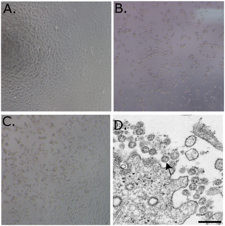
Figure 1.
(A–C) 10X phase contrast images of vero monolayers at 3 days post-inoculation. Panels shown are (A) mock, (B) nasopharyngeal, and (C) oropharyngeal. (D) Electron microscopic image of the viral isolate showing extracellular spherical particles with cross sections through the nucleocapsids, seen as black dots.

