
Figure 2: Reactivity of control and SARS-CoV-2 convalescent sera to different spike antigens.
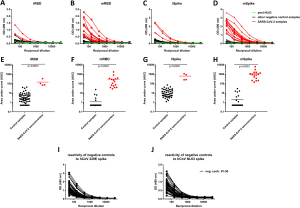
A-D Reactivity to insect cell derived RBD (iRBD), mammalian cell derived RBD (mRBD), insect cell derived soluble spike protein (iSpike) and mammalian cell derived soluble spike protein (mSpike). Sera from SARS-CoV-2 infected individuals are shown in red. One sample, shown in green, is a convalescent serum sample post NL63 infection. E-F shows data from the same experiment but graphed as area under the curve (AUC) to get a better quantitative impression. The n for the control samples is 50 except for the iRBD where it is 59. Statistics were performed using an unpaired two-tailed student’s t-test in Graphpad Prism. I-G shows reactivity of the 50 negative control samples from A-F against spike protein from human coronaviruses 229E and NL63.