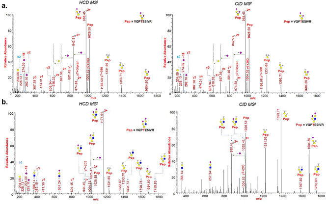
Fig. 7.

HCD and CID MS/MS spectra showing glycan neutral losses, oxonium ions and peptide fragments of (A) representative O-Glycopeptide 320VQPTESIVR328 with core 1 type GalNAcGalNeuAc2 glycan detected on site Thr323 of spike protein subunit S1; (B) representative O-Glycopeptide 320VQPTESIVR328 with core 2 type GalNAcGalNeuAc(GlcNAcGalNeuAc) glycan detected on site Thr323 of spike protein subunit S1. Monosaccharide symbols follow the SNFG system (Varki et al. 2015).
