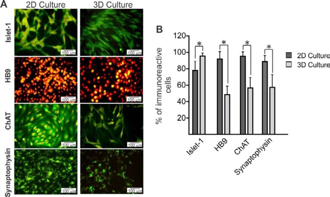
Figure 3.
Immunofluorescence studies were performed to investigate the expression of the motor neuron markers in two different groups. A) MNLCs were immunostained with primary antibodies; islet-1, HB9, ChAT, and synaptophysin. Markers are shown in green and the cell nuclei (counterstained with PI) are shown in red. B) Quantitative data of positive cells in 2D and 3D groups. Our results showed that islet-1 significantly increased in 3D compared with the 2D group. The higher expression of HB9, ChAT, and synaptophysin was observed in 2D more than in 3D. Data are presented as mean±SD. *P<0.05

