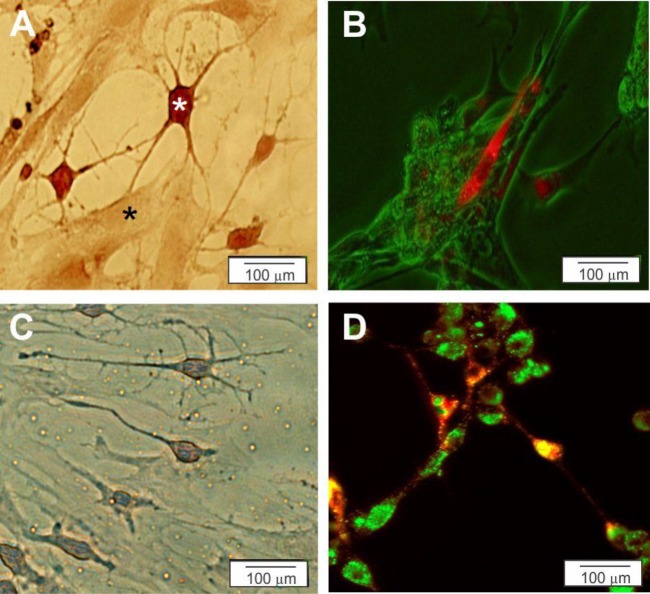
Figure 4.
Characterization of the MNLCs was assessed by co-culturing with myotubes (C2C12) on 2D (A and B) and 3D (C and D) culture plates. A and C) Myotubes co-cultured with MNLCs was stained by Cresyl violet on 2D (A) and 3D (C) culture plates (black star is Myotubes and white star is MNLCs). B and D) Myotubes were stained with PKh67 (green) and co-cultured with the MNLCs were stained with PKh26 (red) on 2D (B) and 3D (D) culture plates

