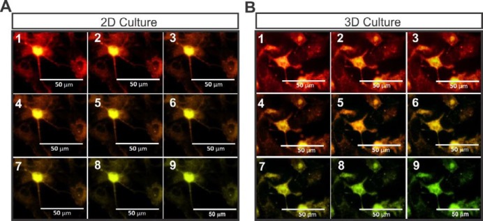
Figure 7.
The staining of the MNLCs with voltage-sensitive dye (RH795) followed by their stimulation on 2D and 3D culture systems. A shows the membrane depolarization and repolarization of the MNLCs with change of color in 2D, and B represents the same field photographed serially demonstrated membrane action potential with same mechanism on 3D culture plates

