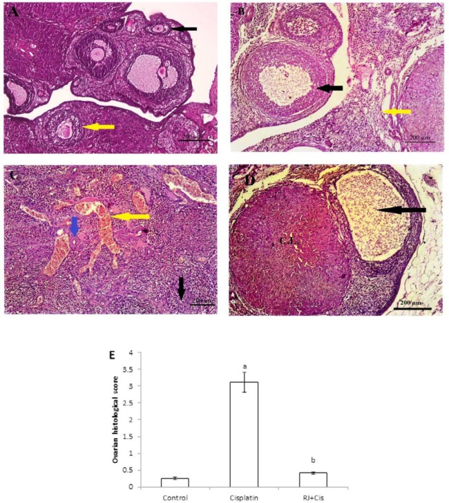
Figure 1.
Representative photomicrographs of H&E-stained sections of rat ovaries in each experimental group
A) Light microscopy of the ovarian tissue with developing follicles in many different stages in the control group. B) Cisplatin group showed a severe histopathologic injury than in other groups, follicular degeneration (black arrow), interstitial edema (yellow arrow). C) A significant marked vascular congestion (yellow arrow), hyalinosis (blue arrow) as well as stromal edema (black arrow) in cisplatin group when compared to the control group. D) Administration of Royal jelly (RJ) with cisplatin in RJ+Cis. group restored the normal structure of the ovarian tissue with no significant difference from control group. E) Quantification of histopathological scores in the ovarian sections from the experimental groups where data were expressed as means±SD, a indicates a significant difference when compared to control group at P˂0.05 and b: indicates a significant difference when compared to cisplatin group (P˂0.05)

