
Fig. 2.4. Early studies of brain glycogen.
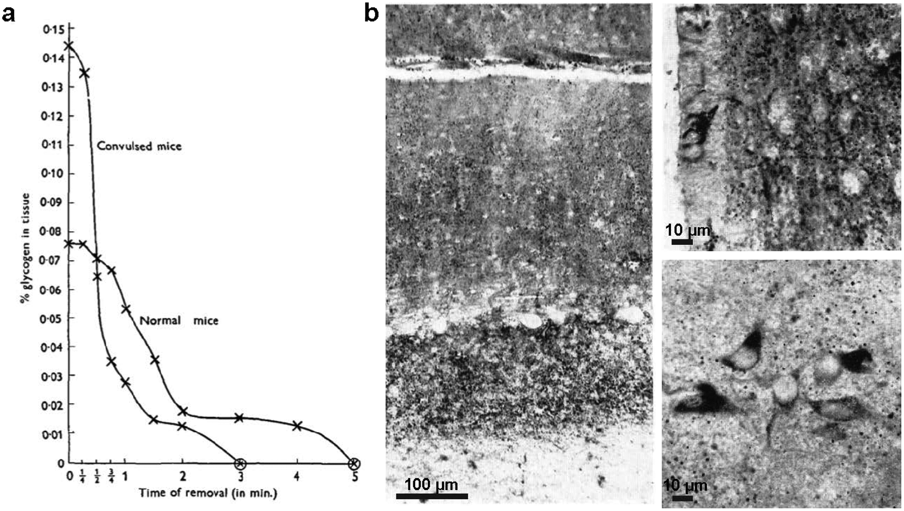
(a) Rapid glycogen loss following extraction from brain of normal and convulsed mice; glycogen was measured using a modification of the method by Kerr (1938) (from Chance and Yaxley 1950). Copyright © Company of Biologists, Ltd. Used with permission. (b) Staining of glycogen (dark stain) in the perfused rabbit brain using the lead-tetra-acetate-Schiff method, a modification of PAS (Shimizu and Kumamoto 1952). The identity of stained regions as glycogen was confirmed by salivary digestion of comparable sections. Left: glycogen is abundant in the granular and molecular layers of the cerebellum, but Purkinje neurons lack glycogen. Upper right: ependymal cells of the hypothalamus lining the third ventricle show intense staining for glycogen, and granules are abundant in the neuropil, but nerve cells lack glycogen in this region. Lower right: some small nerve cells in the lateral hypothalamic nucleus appear to contain glycogen. Scale bars have been approximated based on magnification. Copyright © 2004 John Wiley and Sons, Inc. Used with permission.