
Fig. 2.6. Structure and synthesis of polyglucans.
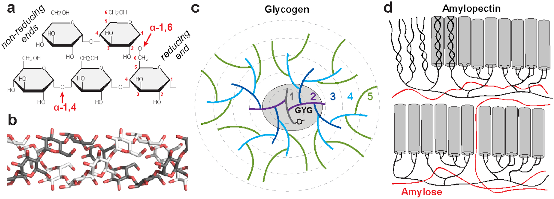
(a) Starch and glycogen contain linear chains of glucose joined by α−1,4 glycosidic linkages, and α−1,6 glycosidic linkages constitute the branch points. The reducing ends (containing an aldehyde group) are oriented near the interior of the polysaccharide molecule, while metabolic enzymes work on the nonreducing ends, which are oriented outward. (b) The α conformation of α−1,4 linked polyglucans gives them a propensity to twist, and long unbranched chains can form single or double helices; a model of a double helix is shown. (c) A model of the first five tiers of the glycogen particle according to the Whelan model. A tyrosine residue of glycogenin (GYG) is shown, which is covalently linked to the glucan chain making up the first tier. (d) A recent model for starch structure according to (Bertoft 2017). In amylopectin, the branched component of starch, branching is clustered, and the long linear chains form double helices (grey cylinders) that make up the crystalline regions of starch. Amylose, the unbranched component of starch, is believed to occupy areas around the amylopectin molecules.