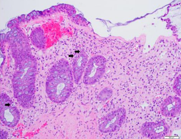
Figure 1.

Colonic tissue with features of graft-versus-host disease. Histologic section of a colonic biopsy shows extensive crypt cell apoptosis, crypt destruction and dropout with focal mucosal necrosis and epithelial denudation. Black arrows indicate most prominent single-cell crypt apoptosis (hematoxylin-eosin, original magnification, × 200).
