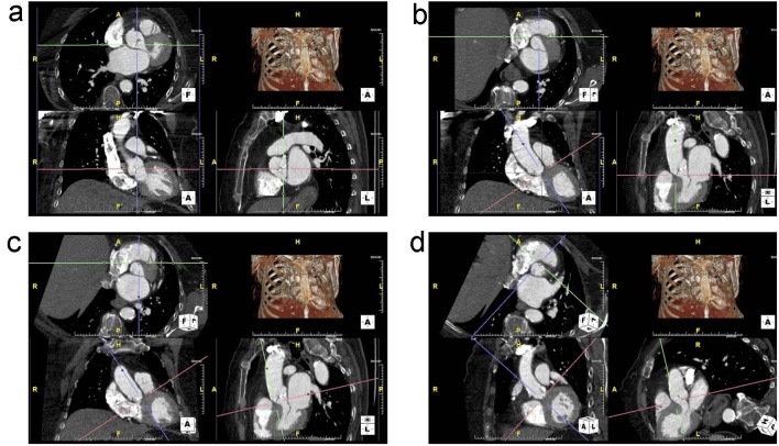
Figure 2.
(a) Unaligned view of the aortic annulus that is not in plane. The top-left is the axial view, bottom-left is the transverse view and the bottom-right is the sagittal view. (b) The right coronary cusp (RCC) is now visible in the axial view by adjusting the red axes. The green and purple axes are then centered on the insertion of the RCC. (c) The non-coronary cusp (NCC) insertion is aligned by adjusting the red axes in the sagittal image to bring it into plane with the RCC insertion in the axial view. (d) The purple axes are adjusted to run through both RCC and NCC insertions. Then, the red axes on the transverse view are adjusted to bring in the insertion of the left coronary cusp (LCC) into the same plane as the RCC and NCC.

