. 2020 Apr 27;9:e54568. doi: 10.7554/eLife.54568

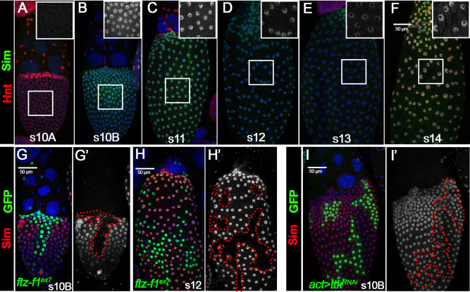
Figure 5. Ftz-f1 promotes Sim expression in stage-10B follicle cells.

(A–F) The expression of Sim protein in late oogenesis. Sim protein is detected by anti-Sim antibody shown in green. Hnt expression (shown in red) is used to mark stage-10A (A) and stage-14 (F) follicles. The insets are higher magnification of Sim expression in squared areas. All images from A-F are acquired using the same microscopic settings. (G–H) Sim expression (red in G,H and white in G’,H’) in stage-10B (G) and stage-12 (H) egg chambers with ftzf1ex7 mutant clones (marked by green GFP and outlined by dashed lines). (I) Sim expression (red in I and white in I’) in stage-10B egg chambers with flip-out Gal4 clones (marked by green GFP and outlined by dashed line) overexpressing ttkRNAi. Nuclei are marked by DAPI in blue.

Figure 5.

Figure 5—figure supplement 1. Sim is detected in stalk follicle cells.
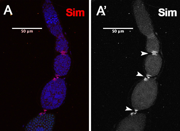
Figure 5—figure supplement 1.

(A) Sim protein (red in A and white in A’) is detected in stalk follicle cells that connects two adjacent egg chambers in the ovariole. White arrowheads point to stalk follicle cells. Nuclei are marked by DAPI in blue.
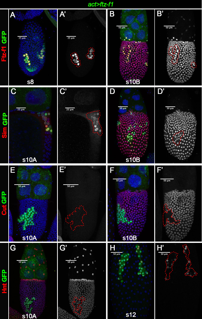
Figure 5—figure supplement 2. Overexpression of ftz-f1 induces premature Sim expression at stage 10A and disrupts follicle cell transition into stage 10B.

Figure 5—figure supplement 2.

Flip-out Gal4 clones with ftz-f1 overexpression are marked by green GFP and outlined in red. (A–B) Ftz-f1 (red in A,B and white in A’,B’) was overexpressed in flip-out Gal4 clones in stage-8 egg chambers (A) and stage-10B egg chambers (B). (C–D) Sim (red in C,D and white in C’,D’) is prematurely expressed in flip-out Gal4 clones at stage 10A (C) and continues to be expressed at stage 10B (D). (E–F) Cut (red in E,F and white in E’,F’) is neither detected in flip-out Gal4 clones at stage 10A (E) nor at stage 10B (F). (G–H) Hnt (red in G,H and white in G’,H’) is properly expressed in stage-10A clones (G), and is continuously expressed in clones cells at stage 12 (H). Nuclei are marked by DAPI in blue.