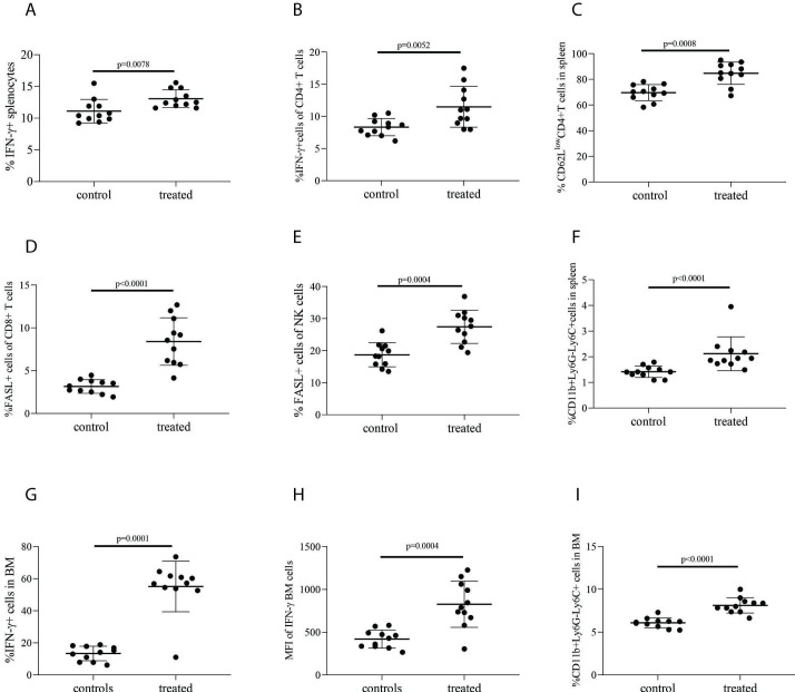
Figure 7.
Impact of inhibiting CD39, CD73, and A2AR in vivo on immune cells. C57BlKalwRij myeloma-bearing mice (n=11) were treated with POM-1, CD73 antibody, and A2AR inhibitor as described in figure 6 and in the Methods section, or saline as control (n=11). Mice were killed on day 15 after tumor injection. Cells from spleen and BM were collected for FACS analysis. Graphs show (A) the percentage of IFN-γ expressing cells in spleen; (B) the percentage of CD4+ T cells from spleen expressing IFN-γ; (C) the percentage of activated CD4+ T cells (CD62LlowCD4+) in spleen; (D) the percentage of FasL+ cells of CD8+ T subsets in spleen; (E) the percentage of FasL+ cells of total NK (CD3−NK1.1+) cells in spleen; (F) the percentage of monocytes (CD11b+Ly6G−Ly6C+) in spleen; (G) the percentage of IFN-γ expressing cells from BM; (H) the MFI of IFN-γ in BM; and (I) the percentage of monocytes (CD11b+Ly6G−Ly6C+) in BM. Results are expressed as mean and SD. Statistical differences were calculated using Mann–Whitney U test. A2AR, adenosine receptor A2A; BM, bone marrow; MFI, median fluorescence intensity.

