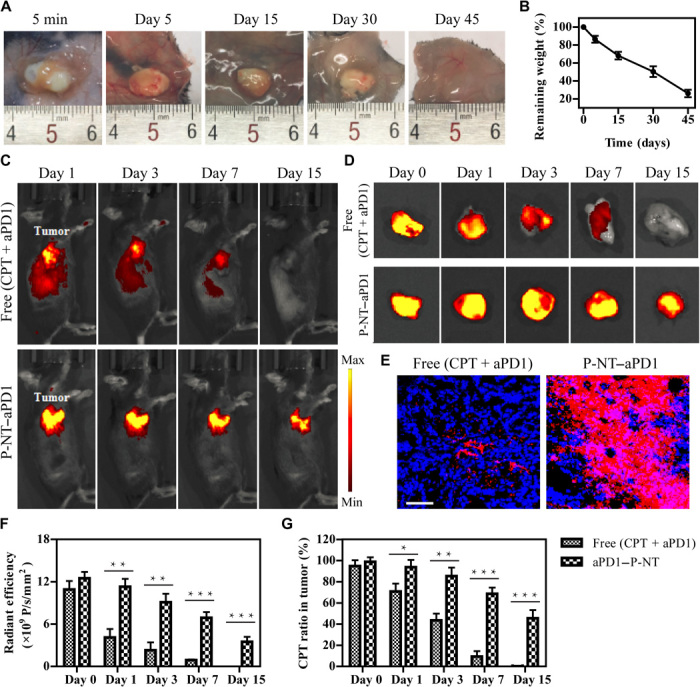
Fig. 2. P-NT–aPD1 hydrogel enhances local retention and prolongs in vivo release of aPD1.

(A) In vivo gel formation and retention after subcutaneous injection of P-NT solution in the back of C57BL/6 mice. (B) In vivo degradation profile of the P-NT hydrogel over time, as determined by the mass loss method. (C) Fluorescence IVIS imaging of the local retention and distribution of aPD1-Cy5.5 in mice, administered in solution form and with the P-NT hydrogel. Experiments were repeated three times. (D) Fluorescence imaging of tumor tissues and (E) tumor sections of GL-261 brain tumor–bearing mice after tumoral injections of free (CPT + aPD1) or P-NT–aPD1. Red, Cy5.5-labeled aPD1; blue, 4′,6-diamidino-2-phenylindole–stained nuclei. Scale bar, 200 μm. (F) Quantification of the in vivo retention profile of Cy5.5-aPD1 and (G) CPT. Statistical significance was calculated using a two-sided unpaired t test. Data are given as means ± SD (n = 3). *P ≤ 0.05, **P ≤ 0.01, and ***P ≤ 0.001. Photo credit: Feihu Wang, Johns Hopkins University.
