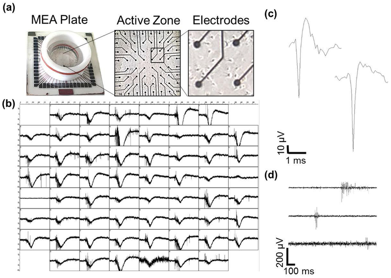
Figure 1.
Spontaneous activity from rat hippocampal neural networks. (a) An MEA (left) and differential interference contrast (DIC) images of a DIV7 culture of hippocampal neurons plated on the MEA at low (center) and high (right) magnification. (b) Network electrical activity from all 60 electrodes of the MEA at DIV14. Each box corresponds to one second of activity (x-axis) with a voltage range of ± 100 μV (y-axis). (c) Representative 3-ms spike traces from a single electrode during baseline. (d) Three representative 1-second traces of filtered activity from an MEA. Within a single electrode, a wide range of different types of bursts is observed, including those with long, intermediate or short duration.

