Fig. 2.
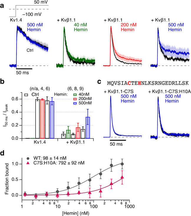
Heme sensitivity of Kvβ1.1-mediated inactivation. a Mean normalized inside-out patch-clamp current traces from HEK293t cells expressing Kv1.4 channels alone (left) or with coexpression of Kvβ1.1 by depolarization steps to 50 mV from a holding potential of − 100 mV. Thick black traces are means before, and the colored traces about 2 min after application of the indicated concentrations of hemin. All solutions additionally contained 200 μM GSH. Shading indicates SEM. For n, see panel (b). b Fraction of non-inactivated current after 50 ms depolarization for Kv1.4 and with coexpression of Kvβ1.1. Data are means ± SEM, n in parentheses. c N-terminal protein sequence of Kvβ1.1 (top). Current traces as in (a) for Kvβ1.1 mutants C7S and C7S:H10A (bottom). Measurements were performed at pH 7.9 to eliminate N-type inactivation endogenous to Kv1.4. d Microscale thermophoresis of the Kvβ1.1 N-terminal domain. Binding curves for interaction of Kvβ1.1 1–140 fused to MBP (gray circles) and mutant C7S:H10A (magenta triangles) as a function of hemin concentration, normalized to the WT data at the highest concentration of hemin. Data are means ± SEM (n = 3; 2 protein preparations)

