Fig. 3.

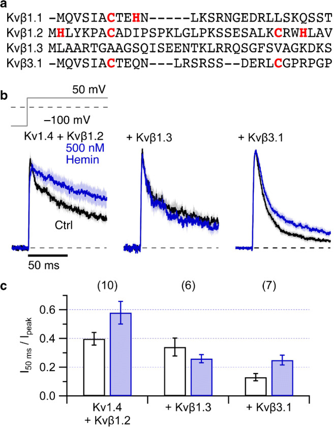
Kv1.4 coexpression with Kvβ1.2, Kvβ1.3, and Kvβ3.1. a N-terminal protein sequence alignment of the Kvβ1 splice variants and Kvβ3.1. b Mean normalized inside-out patch-clamp current traces from HEK293t cells expressing Kv1.4 channels with Kvβ1.2 (left), Kvβ1.3 (center), or Kvβ3.1 (right) by depolarization steps to 50 mV from a holding potential of − 100 mV. Thick black traces are means before and the colored traces about 2 min after application of 500 nM hemin (blue). All solutions additionally contained 200 μM GSH. Shading indicates SEM. For n, see panel (c). Measurements were performed at pH 7.9 to eliminate N-type inactivation endogenous to Kv1.4. c Fraction of non-inactivated current after 50 ms depolarization for Kv1.4 with coexpression of the indicated Kvβ subunits. Data are means ± SEM, n in parentheses
