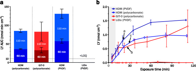
Fig. 3.
(a) LY exposure in receiver with HDM (blue) or LiDo membranes (red), subjected to digestion of a type IIIB medium-chain LBF by porcine pancreatin. Filter support material was either polycarbonate or PVDF (polyvinylidene difluoride). Area under the curve (AUC) values are calculated on exposure time of either 60 min (darker) or 110 min (lighter). Bars reaching above the horizontal dotted line indicate a major loss of integrity. (b) Corresponding LY mass transfer curves for HDM (blue), LiDo or GIT-0 (red), on polycarbonate (circles) or PVDF (triangles) filter support. Arrows point to the time points in each system at which AUC ≈ 10 nmol min cm−2, corresponding to a) 21, b) 26, and c) 29 min of LY exposure. The gray shaded field indicates the time before addition of digestion agent (10 min after experiment start).

