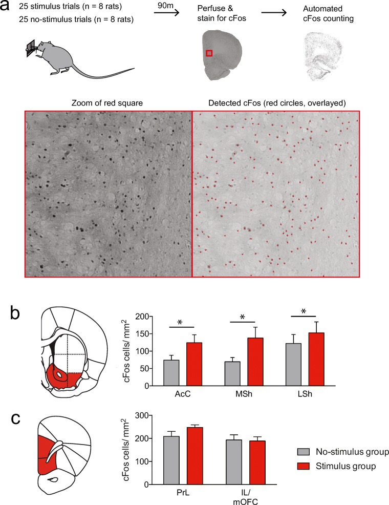
Fig. 4.
Animals that received stimulus trials showed enhanced c-Fos expression in the VS, but not vmPFC, as compared with animals that received no-stimulus trials. a Experimental procedure. Red dots in bottom right figure show the detection of c-Fos by the algorithm. bc-Fos density was enriched across the entire VS after stimulus trials (*P = 0.0153, main effect of group in ANOVA; see Supplementary statistics table in Online Resource 1). AcC, nucleus accumbens core; MSh, medial shell of the nucleus accumbens; LSh, lateral shell of the nucleus accumbens. c Stimulus trials did not evoke changes in c-Fos density in the vmPFC (See Supplementary statistics table in Online Resource 1). PrL, prelimbic cortex; IL, infralimbic cortex; mOFC, medial orbitofrontal cortex

