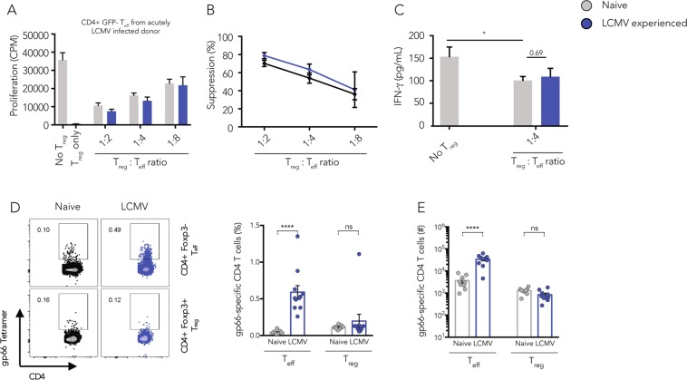
Figure 1.
Infection-experienced Tregs show trend for higher in vitro suppressive capacity. CD4+GFP+ Tregs were sorted from naive or LCMV-experienced Foxp3-GFP reporter mice and co-cultured with CD4+GFP- effector cells isolated from acutely LCMV infected donor mice in the presence of soluble anti-CD3 (1 µg/ml) and irradiated splenic APCs isolated from naive mice for 2 days, then [3H]thymidine was added for 18–22 h and [3H]thymidine incorporation was quantified to determine proliferation. Target cell proliferation (A) and calculated Treg-mediated suppression (B) are shown. (C) The concentration of IFN-γ in the suppression assay supernatants harvested after 48 h of co-culture was determined by cytometric bead assay. (Mean ± SD; biological replicates: no Tregs, naive Tregs, memory Tregs n = 6; 2 independent experiments) (one way ANOVA and multiple comparisons test, *p < 0.05, **p < 0.01, ***p < 0.001). (D) Splenic T cells from naive or LCMV-experienced mice (>30 days post infection) were stained using gp66 tetramers to detect LCMV-specific CD4+Foxp3+ Tregs or CD4+Foxp3- effector T cells. Frequencies and absolute numbers are shown. (Mean ± SD; biological replicates: naive n = 8, LCMV-experienced n = 9; 3 independent experiments) (t Test, ****p < 0.0001).

