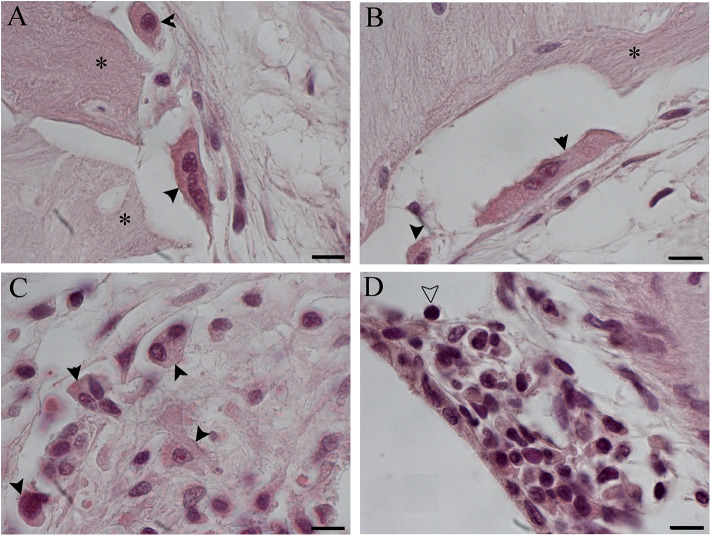
Figure 8.
Foreign body giant cells (FBGCs) in proximity of new bone growth in the outer (A) and inner (B) side of the cochelostomy. Numerous FBGCs are visible also inside the inflammatory reaction around the electrode (C). An inflammatory infiltration is visible near the cochleostomy (D). Hematoxylin–eosin staining. Scale bars, 10 μm. *, new bone; black arrowheads, FGBC; white arrowhead, lymphocyte.

