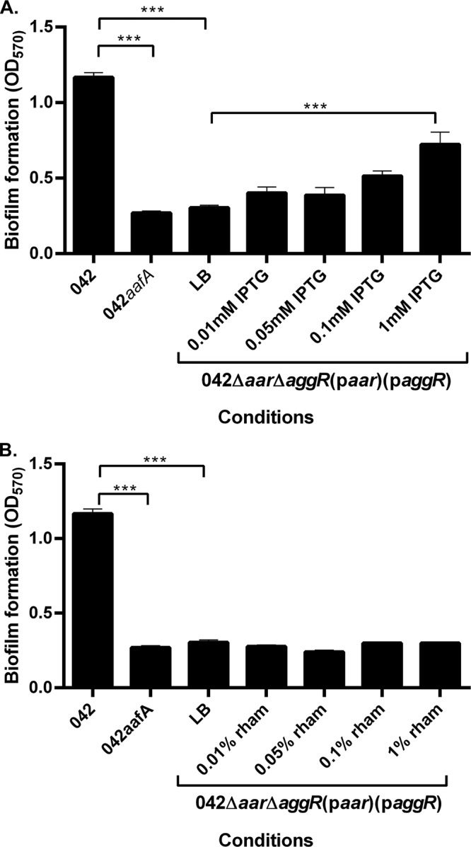
FIG 1.

Biofilm formation in the presence and absence of inducer molecules. (A) Biofilm formation was measured using crystal violet staining after 3 h in 042 and 042ΔaafA in DMEM high glucose and in 042Δaar ΔaggR(paar)(paggR) in LB with varying concentrations of IPTG. (B) Biofilm formation was measured using crystal violet staining after 3 h in 042 and 042ΔaafA in DMEM high glucose and in 042Δaar ΔaggR(paar)(paggR) in LB with varying concentrations of rhamnose. Biofilm data are representative of at least three independent experiments. Asterisks indicate significant differences by ANOVA (*, P < 0.05; **, P < 0.005; ***, P < 0.0005).
