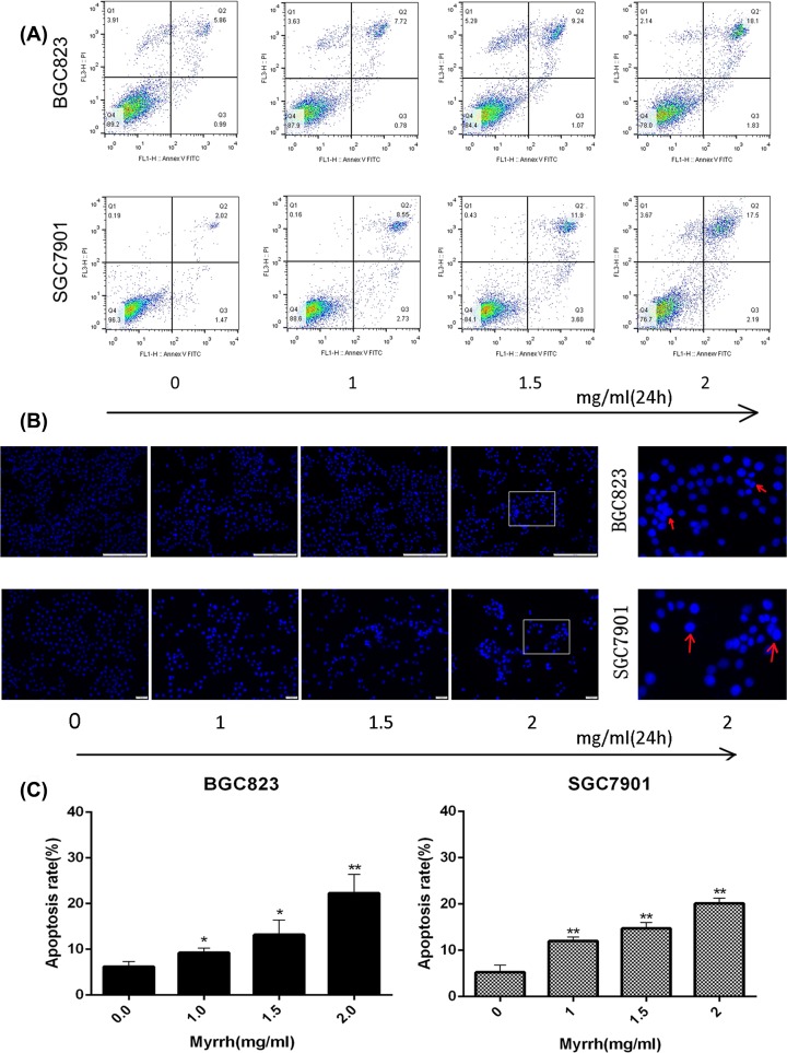
Figure 2. Myrrh induces apoptosis in GC cells.
(A) Flow cytometry-based annexin V-FITC/PI labeling of apoptotic cells. (B) Morphological changes in apoptotic cells were examined by fluorescence microscopy after Hoechst 33342 staining. Morphological changes were seen only in myrrh-treated gastric cancer cells. Myrrrh-treated BGC-823 and SGC-7901 cells clearly showed condensed and fragmented nucleus and emitted bright fluorescence. (C) The histogram represents apoptosis rates. Each data point represents the mean ± SD from three independent experiments; *P<0.05, **P<0.01 versus control (0 mg/ml)

