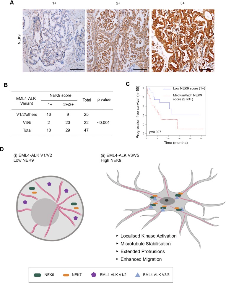
Fig. 8.
Elevated NEK9 expression correlates with EML4–ALK V3 or V5 genotype in NSCLC patient tumours as well as poor overall survival. (A) Representative images of tumour biopsies from NSCLC patients that were processed for immunohistochemistry with NEK9 antibodies (brown) and scored as low (1+), medium (2+) or high (3+) expression. Tissue was also stained with haematoxylin to detect nuclei (blue). Scale bars: 200 μm. (B) NEK9 expression, scored as in A, with respect to the EML4–ALK variant present. (C) Kaplan–Meier plot indicating the progression-free survival of NSCLC patients with EML4–ALK fusions that had low (1+) or medium/high (2+/3+) NEK9 expression (n=32). (D) Schematic model showing rationale for stratification of cancer patients for treatment based on EML4–ALK variant. (i) The majority of tumours expressing EML4–ALK V1 or V2 express low levels of NEK9. In these cells, the EML4–ALK protein neither binds NEK9 nor colocalizes with microtubules, and cells retain a more rounded morphology. (ii) However, the majority of tumours expressing EML4–ALK V3 or V5 express moderate or high levels of NEK9. In these cells, the EML4–ALK protein binds and recruits NEK9 and NEK7 to microtubules leading to localized kinase activity that promotes microtubule stabilization, formation of extended cytoplasmic protrusions and enhanced migration.

