
Fig. 6 |. PEG-4MAL hydrogel promotes HIO engraftment into mucosal wounds.
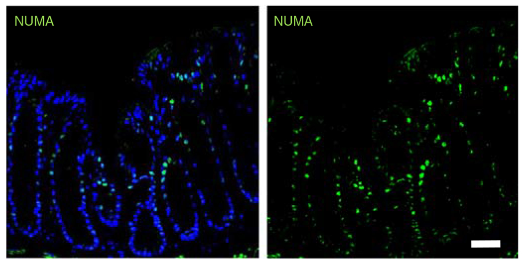
Fluorescence microscopy images of murine colonic tissue at the wound site labeled for human cell nuclei (NUMA, EMD Millipore, cat. no. MAB1281, overnight incubation at 1:100; followed by donkey anti-mouse Alexa Fluor 488, Jackson ImmunoResearch, cat. no. 715-545-150, 1-h incubation at 1:2,000; green) taken 4 weeks post delivery. DAPI (blue; 1:1,000), was used as counterstain. Scale bar, 100 μm. All experiments with mice were performed while following national and institutional guidelines. These data are representative of the results we saw after injection of hydrogels containing HIOs in two independent studies (n = 4 mice per condition, five injections per mouse). Further results from this study are published in ref. 18 Samples were imaged using a Zeiss Axiovert 35 microscope and analyzed with LAS X software (Leica Microsystems).