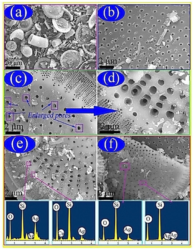
Figure 3.
(a) Illustrates the disc and cylindrical like structures of the raw diatomite powder. This also indicated impurities blocking the pores of the diatomite. (b) After acid treatment, some of the impurities were removed. (c) Morphology of the porous structure was maintained after alkali leaching. (d) Enlargement of (c) indicating where the PEG would disperse the AgNPs in the pores of the purified diatomite. (e,f) Indicates the deposit of the AgNPs on the surface of the structure. The energy dispersive X-ray spectroscopy spectra confirmed the presence of silicon, oxygen, and silver in the composite, while proper AgNP dispersion was confirmed visually [41].

