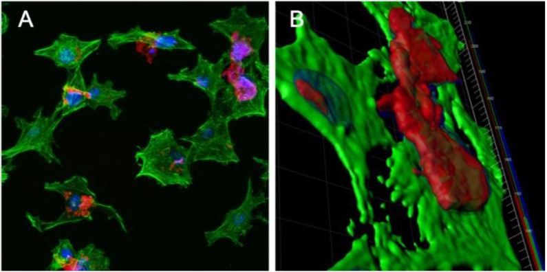
Figure 8.
Confocal microscopy images of IMAC cells treated with 100 μg/mL Nile red-labeled PBSe-GSK3787 particles (PBSe-GSK3787-NR) particles (red) for 48 h, then stained with AlexaFluor 488 Phalloidin (green, cytoskeletons) and 4’,6-diamino-2-phenylindole (DAPI) (blue, nuclei): (A) 2D image showing agglomerates of particles on the cells; (B) 3D rendering of cells showing particles localized at the cell surface and not taken up by the cells.

