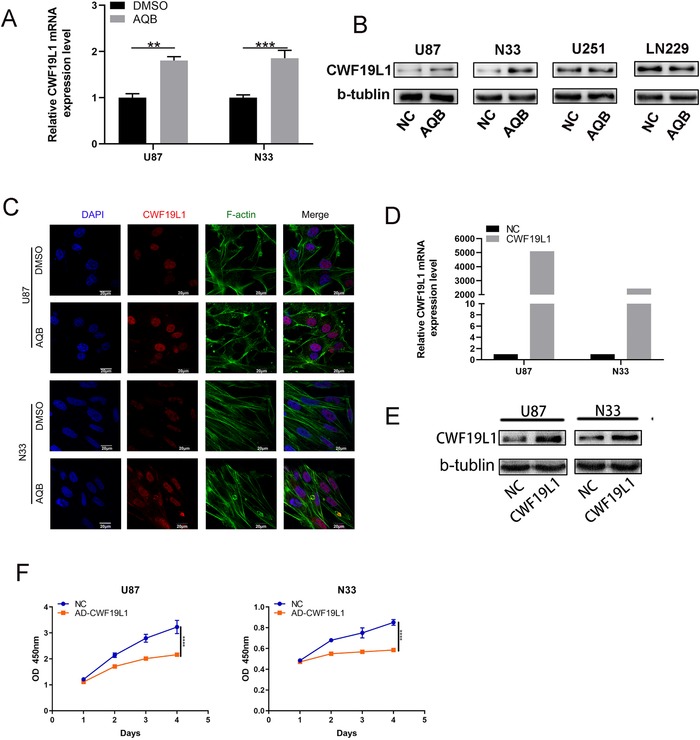
FIGURE 3.

The key role of CWF19L1 in the proliferation of glioma cells. A, The western blot data revealed the upregulation of CWF19L1 protein levels in U87 and N33 cells, while CWF19L1 expression levels were not significantly different in U251 and LN229. B, The upregulation of CWF19L1 mRNA levels in U87 and N33 cells after AQB treatment (* P < .05, ** P < .01, *** P < .001). C, The confocal microscopy revealed that AQB upregulated CWF19L1 and remodeled the actin (F‐actin) cytoskeleton. DAPI was used to stain the nuclei. Bar, 20 um. E, CWF19L1 expression increased in the overexpression group. F, Detection of the proliferation of U87 cells by CCK‐8 assay after the transfection of the CWF19L1 plasmid
