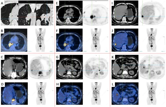
FIGURE 2.

Imaging phenomes of positron emission tomography/computed tomography (PET/CT) 4 months after onset (continuation of Figure 1): Lung MT with multiple systemic metastases, more bone metastases and liver metastases, brain metastasis, left submaxillary lymph node metastasis, spleen and left adrenal gland metastasis. A,B,E,F, PET/CT images of the chest. C, D, and G‐L, PET/CT images of abdomen
