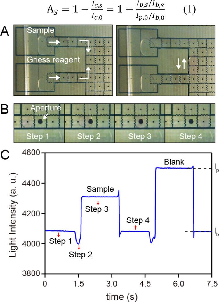
Figure 2.

(A) Photographs of the droplets disposing for nitrite sample and Griess reagent (left) and the droplet-based Griess reaction (right) on the DMF. (B) Photographs of the droplet movements (steps 1–4) on the sensing electrode. For steps 1 and 4, the droplet is outside the sensing electrode. For step 2, part of the droplet enters the sensing electrode. For step 3, the whole droplet moves to and is stopped at the center of sensing electrode; except the position labeled as “Aperture”, the other aperture-like structure is just the linkage of each electrode for applying voltage. (C) Light response corresponding to the four steps; definitions of the Ip, Ib is labeled on the light pulse of the blank droplet as an example.
