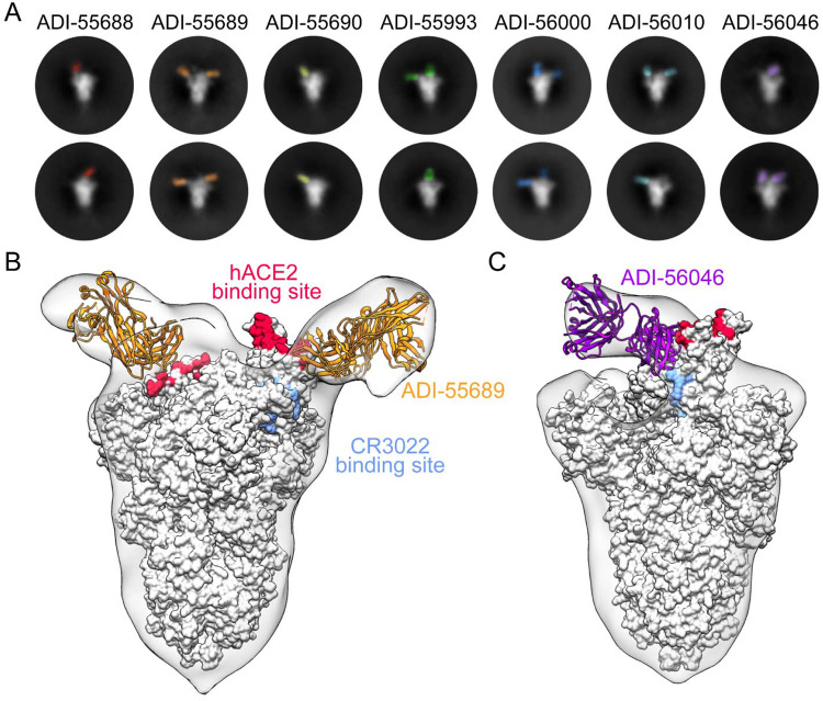
Figure 4.
Structures of cross-neutralizing antibodies bound to SARS-CoV-2 S. (A) Negative-stain EM 2D class averages of SARS-CoV-2 S bound by Fabs of indicated antibodies. The Fabs have been pseudo-colored for ease of visualization. (B-C) 3D reconstructions of Fab:SARS-CoV-2 S complexes are shown in transparent surface representation (light gray) with the structure of the SARS-CoV-2 S trimer docked into the density (white surface). Fabs have been docked into the density and are shown in ribbon representation. S-bound Fabs of ADI-55689 (B) and ADI-56046 (C) are colored in orange and purple, respectively. The hACE2 and CR3022 binding sites on S are shaded in red and light blue, respectively.

