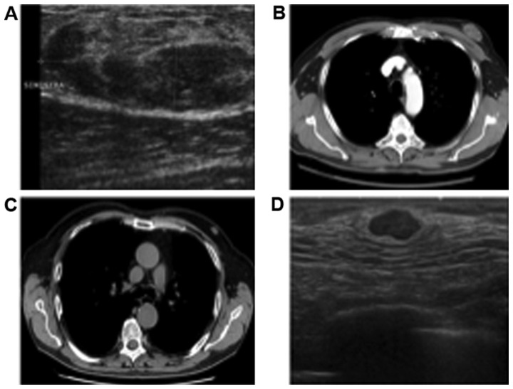
Figure 1.
Radiological examination of mammary myofibroblastoma. The images represent the MFB mass observed in ultrasonography in (A) the first and (D) second patient. Both images show a well-circumscribed, oval, lesion. The axial imaging represents a CT scan in (B) the first and (C) second patient, with both patients showing a breast mass, well circumscribed. MFB, mammary myofibroblastoma; CT, computed tomography.

