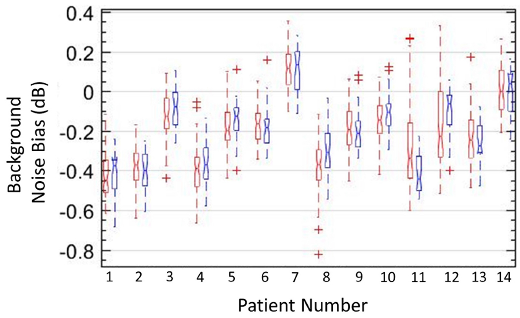
Figure 12:

Box-and-whisker plots of background data obtained from vertical (red) and horizontal (blue) regions of interest in background-suppressed power Doppler images of 14 in vivo cases, with lines indicating the lower, median, and upper quartiles.
